Advanced Technology in Optometry

Contact Us
- Vision Plus - Corporate Office
2520 James Street
Bellingham, WA
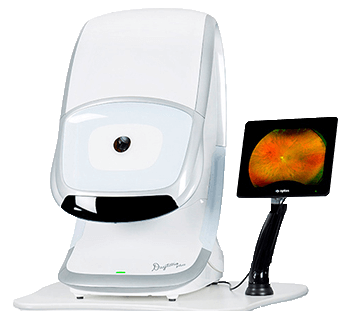
Optomap Retinal Imaging
Now Available At Many Vision Plus Clinics
Your optometrist might need to apply eye drops to dilate your pupils. This can make your eyes a bit blurred and more sensitive to light, which could affect your close vision for up to 5 hours. Most of our offices now have the Optos retinal camera which may allow us to view your eyes without drops.

Corneal Thickness Mapping
Pachymetry also known as corneal thickness mapping is a process used to measure the thickness of the cornea. The two modern technologies available are ultrasound and optical coherence tomography (OCT). Ultrasound uses reflected sound waves to measure corneal thickness where OCT uses reflected light waves to perform the same goal. This technology helps us tailor testing options and treatment results specifically for you. Eye pressure and risks of specific eye conditions are much more likely to be accurate with a known corneal thickness.
Both technologies are available at many of our Vision Plus locations. This testing is necessary for the assessment of glaucoma, LASIK analysis, corneal ectasia diagnosis, and evaluation and management of corneal procedures or dystrophies that might involve swelling.
Corneal Topography
Corneal topography is a non-intrusive medicinal imaging method for mapping the surface shape of the cornea. This innovation takes into consideration the following three dimensions: the exact contact focal point fits, screening for sporadically formed corneas, and enhancing refraction exactness for glasses remedies. Since the cornea is regularly in charge of some 70% of the eye's refractive power, its geology is of basic significance in deciding the nature of vision.
The three-dimensional guide is a profitable indicator to the analyzing optometrist and can aid the analysis and treatment of various conditions; in arranging refractive medical procedure, for example, LASIK and assessment of its outcomes; or in evaluating the attack of contact focal points.
Digital Retinal Imaging & OCT Scans
We utilize front-line advanced imaging innovation to survey your eyes. Many eye illnesses, whenever identified in the early stages, can be dealt with effectively without complete loss of vision. Your retinal Images will be stored electronically. This gives the eye specialist a continuous record on the condition of your retina. This is critical in helping your optometrist recognize and quantify any progressions to your retina each time your eyes are examined. The same number of eye conditions, for example, glaucoma, diabetic retinopathy, and macular degeneration are analyzed by distinguishing changes after some time. The upsides of advanced imaging include:
- Quick, safe, non-invasive and painless analysis
- Detailed images of your retina and sub-surface of your eyes
- Instant, direct imaging of the form and structure of eye tissue
- Extremely high-quality Image resolution
- Maintaining eye-safety near-infra-red light
- No patient prep required

Humphrey Visual
Field Perimeter
A visual field test measuring the range of your peripheral or “side” vision to assess whether you have any blind spots (scotomas), peripheral vision loss or visual field abnormalities. You will be required to position your head on a chin rest and look ahead. Lights are flashed in your peripheral vision, and you press a button whenever you see the light. Each eye is tested separately, and the entire test takes 15 to 25 minutes. A computerized map of your visual field is created to identify if and where you have any deficiencies.
Anterior
Segment Imaging
In today’s advanced eye care practices, photo documentation is essential for proper patient care. Slit lamp imaging provides documentation for corneal disease and is utilized as a quality patient education tool. Its primary use is for the detection and quantification of angle closure as it related to glaucoma diagnosis and care. As one of the most rapidly advancing fields in ophthalmology, anterior segment imaging provides optimum care for corneal disease, refractive surgery, contact lens care, and documentation for referrals.
Optical Coherence
Tomography (OCT)
An Optical Coherence Tomography scan (commonly referred to as an OCT scan) is the latest advancement in imaging technology. OCT uses harmless light to create an image of the layers of the retina. These scanning light rays analyze the layers of the retina and optic nerve for any signs of eye disease. Through an OCT scan, detailed images are created, which is revolutionizing early detection and treatment of eye conditions such as wet and dry age-related macular degeneration, glaucoma, retinal detachment, and diabetic retinopathy.
